What Is Scleral Buckling Surgery?
Scleral buckling surgery is a procedure to repair a retinal detachment. It consists of inserting silicone material to return this tissue to its original state.
There are other alternatives, although this is one of the most commonly used by ophthalmologists. Below you will find the most important details about this procedure. So keep reading!
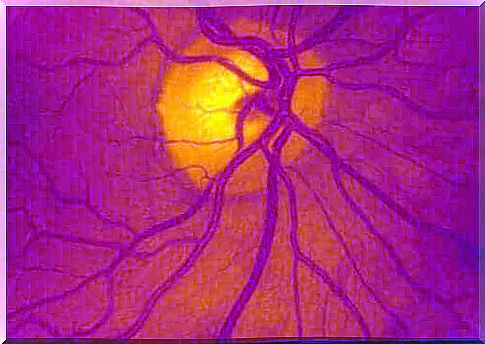
Why does the retina detach?

This structure looks like a thin layer at the back of the eye, but is quite complex microscopically. It is thanks to the retina that we can see images clearly and therefore any damage leaves permanent consequences.
There are many mechanisms involved in retinal detachment and there is an accumulation of fluid (called vitreous humor) behind the structure in each of them. This leads to the gradual loss of blood supply and subsequent death of the tissue.
Detachment can occur spontaneously in some people with risk factors. According to the Mayo Clinic publication (Spanish link), the main ones include:
- Be older than 50 years.
- have diabetes mellitus.
- Have a personal or family history of retinal detachment.
Finally, have had a previous ophthalmic procedure.
How does scleral buckling surgery work?
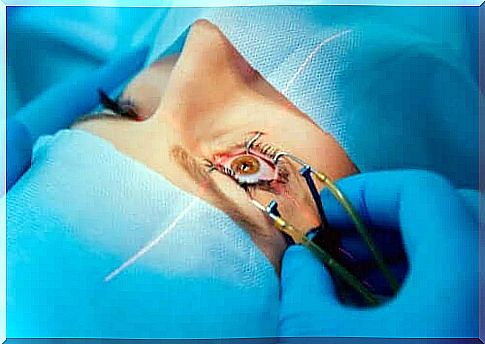
According to a Texas Retina Associates publication, scleral buckling surgery involves the introduction of a silicone band or sponge near the sclera. The last structure is the white part that is visible in the eyes, around the iris.
This causes a slight increase in pressure and displacement of the intraocular structures to return the retina to its original position. The procedure requires several incisions on the outer surface of the eye to insert the material.
The attending physician may apply some additional techniques to prevent recurrence of the disease. The most commonly used are laser photocoagulation and cryopexy. In both cases, scar tissue seals the breaks in the damaged tissues.
Preparation
As with all surgical procedures, there is usually a short preoperative phase. The preparatory phase in this treatment includes the following:
- Stay in bed to avoid sudden movements while you wait for the procedure to begin.
- The doctor or nurse may trim the eyelashes slightly and put in some eye drops to widen the pupil.
- Administration of local or regional anesthesia – this depends on the patient and clinical severity.
How does the procedure work?
Depending on the severity of the retinal detachment, scleral buckling surgery can take at least one to two hours. The general steps in scleral buckling surgery are:
- Make an incision in the white of your eye.
- Then place the silicone material
- A suture of the latter to prevent shifting.
- Finally, there is the drainage of the fluid that has accumulated behind the retina.
It is also possible to perform the complementary procedures from the previous section, even though these procedures all have their own technique.
Expectations and recovery after scleral buckling surgery
The effectiveness of this procedure is usually excellent, even though there are some complications (we’ll list them in the next section). There will be somatic pain during the first days of recovery along with redness and constant tearing. However, these symptoms will subside as the days go by. In addition, the doctor can give other indications, such as:
- Rest, but not always lying down.
- Use an eye patch after the procedure.
- Administer antibiotics regularly.
- Taking pain relievers such as ibuprofen.
- Do not drive or lift heavy objects.
- Do not travel by plane.
- Wearing sunglasses.
As the weeks go by, you should consult your treating physician to verify the effectiveness of the procedure. He can also tell you which measures are and are not necessary.
What are the risks of surgery?
Scleral buckling surgery has some risks because it involves the eyeball. One should pay attention to the warning signs. There are times when the procedure can cause complications in the first few weeks, although this is rare. The main conditions or complications include:
- Infections
- Cataracts
- Glaucoma
- New retinal detachment
Therefore, in case of loss of visual acuity or if you see light or foreign objects, consult an ophthalmologist. Swelling around the eye and increased pain are other warning signs.
Scleral buckling surgery is an effective emergency option
Retinal detachment is a medical emergency that can cause permanent blindness. Consult an ophthalmologist as soon as possible if you notice the above symptoms. Finally, there are other effective procedures, such as pars plana vitrectomy, but scleral buckling surgery remains the first choice for many specialists.